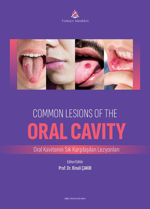
Oral mucosa lesions are the third-most common oral pathology after caries and periodontal diseases. Oral mucosa lesions can be found in each site of the oral mucosa. Clinicians encounter various oral lesions in everyday practice. Clinical findings give important clues about the etiology and diagnosis of the lesion. Oral lesions can arise from a range of different aetiologies: infective, idiopathic, inflammatory, reactive and neoplastic changes. Hovewer, many lesions have known etiopathology and can be treated and eliminated easily and effectively. Knowledge of clinical characteristics such as size, location, surface morphology, color, pain, and duration is helpful in establishing a diagnosis. Oral mucosa lesions are diagnosed worldwide in any population, age or gender, but in varied prevalence. A clinician must obtain a thorough clinical history and have adequate knowledge of the signs and symptoms, such as the location of the oral mucosal lesion and its size, colour and morphology to make a proper diagnosis. Early diagnosis and appropriate approaches of oral lesions will reveal positive results in the prognosis of the diseases and the quality of life of the patients. Clinicians play a central role in early detection of these oral pathologies. The success of a rapid and correct diagnosis of oral mucosal lesions is of great advantage for the patient, ensuring timely management and correct treatment procedures. Most importantly, the timely detection and exclusion of malignant lesions upon a routine dental oral examination provide the clinician and patient with maximum success in treatment opportunities. Large-scale, population-based screening studies have identified the most common oral lesions as candidiasis, recurrent herpes labialis, recurrent aphthous stomatitis, mucocele, fibroma, mandibular and palatal tori, pyogenic granuloma, erythema migrans, hairy tongue, lichen planus, and leukoplakia. The rationale for choosing the lesions included in the present book is that they are the most common oral mucosal diseases that general practitioners experience in their practice. This book has been designed to review common oral lesions that clinicians are faced with in everyday practice and provide an overview, and to guide the clinician to correct and efficient approach in the diagnosis of such lesions.
I believe that it will be useful as a reference book for dentists, medical doctors and other researchers in the field of health.
I would like to thank all the authors who contributed to the writing of this book.
Prof. Dr. Binali ÇAKIR
Atatürk University Faculty of Dentistry,
Department of Oral and Maxillofacial Radiology, Erzurum, Türkiye
Bölümler
Understanding Recurrent Aphthous Stomatitis: Comprehensive Insights Into Causes, Symptoms, and Innovations in Treatment
Muhammed Enes Naralan
Oral Candidiasis
Mustafa Taha Güller
Soft Tissue Calcifications
Hatice Güller, Muhammed Akif Sümbüllü
Oral Leukoplakia
Gülsüm Akkaya
Erythroplakia
Mehmet Akyüz, Güldane Mağat
Herpes Simplex Infection
Esin Akol Görgün, Ahmet Tohumcu
Lichen Planus
Ayşegül Öndeş, Anzel Toprak Bişkin, Esin Akol Görgün
Reactive Hyperplasias of the Oral Cavity
Ahmet Tohumcu, Esin Akol Görgün
Oral Manifestations of Bacterial Infections
Fatma Nur Yozgat İlbaş
Tongue Diseases
Fatma Ceren, Taha Emre Köse


