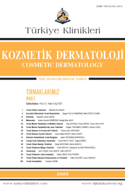
Benign Tumors of Nail Unit
Seher BOSTANCIa, Zehra AKKAYAb
aAnkara Üniversitesi Tıp Fakültesi, Deri ve Zührevi Hastalıkları ABD, Ankara, TÜRKİYE
bAnkara Üniversitesi Tıp Fakültesi, Radyoloji ABD, Ankara, TÜRKİYE
Bostancı S, Akkaya Z. Tırnak ünitesi benign tümörleri. Koçyiğit P, editör. Tırnaklarımız. 1. Baskı. Ankara: Türkiye Klinikleri; 2020. p.60- 72.
ABSTRACT
Because of the anatomical features of nail unit, the clinical differential diagnosis of benign tumors of this specific area is difficult. As the nail plate covers the lesions the clinical appearance of tumors is modified. We reviewed benign tumors and tumor-like lesions of nail unit in relation to the clinical presentation, anatomic location, histopathological features, radiological approach and differential diagnoses.
Keywords: Nails; benign tumors
Kaynak Göster
Referanslar
- Thomas l, Zook Eg, Haneke E, Drape Jl, Baran R. Tumors of the nail apparatus and adjacent tissues. In: Baran R, Berker DAR, Holzberg M, Thomas l, eds. Baran & Dawber's Diseases of the Nails and Their Management. 4th ed. Wiley-Blackwell. 2012. p.637-743. [Crossref]
- Kimbauer R, lenz P. Human Papillomaviruses, section 12 (79). In: Bolognia Jl, Schaffer J, Cerroni l, eds. Dermatology. 4th ed. Elsevier SMT E Books; 2018. p.1383-1399.
- Haneke E. Nail surgery. Clin Dermatol. 2013;31(5):516-25. [Crossref] [PubMed]
- McCown H, Thiers B, Cook J, Ackers S. global nail dystrophy associated with HPV type S7 infection. Br J Dermatol. 1999;141(4): 731-5. [Crossref] [PubMed]
- Wortsman X, Sazunic I, Jemec gB. Sonography of plantar warts: role in diagnosis and treatment. J ultrasound Med. 2009;28(6):787- 93. [Crossref] [PubMed]
- Sorour NE, Elesawy FM, Abdou Ag, Abdelazeem SE, Akl EM. Intralesional injection of vitamin D in verruca vulgaris increases cathelicidin (ll37) expression, therapeutic and immunohistochemical study. J Dermatol Treat. 2020;1-6. [Crossref] [PubMed]
- Zeller J, Friedmann D,Clerici T,Revuz J. The significance of a single periungual fibroma:report of seven cases. Arch Dermatol. 1995;131:1465-6. [Crossref]
- Sezer E, Bridges Ag, Koseoglu D, Yuksek J. Acquired periungual fibrokeratoma developing after acute staphylococcal paronychia. Eur J Dermatol. 2009;19(6):636-7. [Crossref] [PubMed]
- Willard KJ, Cappel MA, Kozin SH, Abzug JM. Benign subungual tumors. J Hand Surg Am. 2012;37(6):1276-86. 10. Kikuchi I, Ishii Y, Inoue S. Acquired periungual fibroma. J Dermatol. 1978;5(5):235-7. [Crossref] [PubMed]
- Haneke E. Intraoperative differential diagnosis of onychomatricoma, Koenen's tumours, and hyperplastic Bowen's disease. J Eur Acad Dermatol Venereol. 1998;11(Suppl):S119. [Crossref]
- Kint A,Baran R,de Keyser H. Acquired(digital) fibrokeratoma. J Am Acad Dermatol.1985;12:816-21. [Crossref] [PubMed]
- Goettmann S, Drape Jl, Idy-Peretti I, Bittoun J, Thelen P, Arrive l, et al. Magnetic resonance imaging: a new tool in the diagnosis of tumours of the nail apparatus. Br J Dermatol. 1994;130(6):701-10. [Crossref] [PubMed]
- Butler ED, Hamill JP, Seipel RS, de lorimier AA. Tumours of the hand. A ten-year survey and report of 437 cases. Am J Surg. 1960;100:293-302. [Crossref] [PubMed]
- Kinoshita Y, Kojima T, Furusato Y. Subungual dermatofibroma of the thumb. J Hand Surg Br. 1996;21(3):408-9. [Crossref] [PubMed]
- Baran R, Perrin CH, Baudet J, Requena l. Clinical and histological patterns of dermatofibromas (true fibromas) of the nail apparatus. Clin Exp Dermatol. 1994;19(1):31-5. [Crossref] [PubMed]
- Mundada P, Becker M, lenoir V, Stefanelli S, Rougemont Al, Beaulieu JY, et al. High resolution MRI of nail tumors and tumor-like conditions. Eur J Radiol. 2019;112:93-105. [Crossref] [PubMed]
- Halteh P, Magro CM, lipner SR. Subungual pleomorphic fibroma: a case report and review of the literature. Dermatol Online J. 2016;22(11):13030/qt9w83z9bq. [Crossref] [PubMed]
- Baran R, Perrin C. localized multinucleate distal subungual keratosis. Br J Dermatol. 1995;133(1):77-82. [Crossref] [PubMed]
- Tosti A, Schneider Sl, Ramirez-Quizon MN, Zaiac M, Miteva M. Clinical, dermoscopic, and pathologic features of onychopapilloma: A review of 47 cases. J Am Acad Dermatol. 2016;74(3):521-6. [Crossref] [PubMed]
- de Berker DA, Perrin C, Baran R. localized longitudinal erythronychia: diagnostic significance and physical explanation. Arch Dermatol. 2004;140(10):1253-7. [Crossref] [PubMed]
- Jellinek NJ. longitudinal erythronychia: suggestions for evaluation and management. J Am Acad Dermatol. 2011;64(1):167. e1-11. [Crossref] [PubMed]
- Miteva M, Fanti PA, Romanelli P, Zaiac M, Tosti A. Onychopapilloma presenting as longitudinal melanonychia. J Am Acad Dermatol. 2012;66(6):e242-3. [Crossref] [PubMed]
- Perrin C, Cannata gE, Bossard C, grill JM, Ambrossetti D, Michiels JF. Onychocytic matricoma presenting as pachymelanonychia longitudinal. A new entity (report of five cases). Am J Dermatopathol. 2012;34(1):54-9. [Crossref] [PubMed]
- Perrin C. Tumors of the nail unit. A review. Part I: acquired localized longitudinal melanonychia and erythronychia. Am J Dermatopathol. 2013;35(6):621-36. [Crossref] [PubMed]
- Perrin C. Tumors of the nail unit. A review. Part II: acquired localized longitudinal pachyonychia and masked nail tumors. Am J Dermatopathol. 2013;35(7):693-712. [Crossref] [PubMed]
- Perrin C, langbein l, Ambrossetti D, Erfan N, Schweizer J, Michiels JF. Onychocytic carcinoma: a new entity. Am J Dermatopathol. 2013;35(6):679-84. [Crossref] [PubMed]
- Perrin C, goettmann S, Baran R. Onychomatricoma: clinical and histopathologic findings in 12 cases. J Am Acad Dermatol. 1998;39(4 Pt 1):560-4. [Crossref] [PubMed]
- Wang l, gao T, Wang g. Nail bed onychomatricoma. J Cutan Pathol. 2014;41(10): 783- 6. [Crossref] [PubMed]
- Perrin C, Cannata gE, langbein l, Ambrossetti D, Coutts M, Balaquer T, et al. Acquired localized longitudinal pachyonychia and onychomatrical tumors: A comparative study to onychomatricomas (5 cases) and onychocytic matricomas (4 cases). Am J Dermatopathol. 2016;38(9):664-71. [Crossref] [PubMed]
- Wortsman X, Wortsman J, Soto R, Saavedra T, Honeyman J, Sazunic I, et al. Benign tumors and pseudotumors of the nail: a novel application of sonography. J ultrasound Med. 2010;29(5):803-16. [Crossref] [PubMed]
- Grigore lE, Baican CI, Botar-Jid C, Rogojan l, letca AF, ungureanu l, et al. Clinico-pathologic, dermoscopic and ultrasound examination of a rare acral tumour involving the nail - case report and review of the literature. Clujul Med. 2016;89(1):160-4. [Crossref] [PubMed] [PMC]
- Oteo-Alvaro A, Meizoso T, Scarpellini A, Ballestin C, Perez-Espejo g. Superficial acral fibromyxoma of the toe, with erosion of the distal phalanx. A clinical report. Arch Orthop Trauma Surg. 2008;128(3):271-4. [Crossref] [PubMed]
- Sundaramurthy N, Parthasarathy J, Mahipathy SR, Durairaj AR. Superficial Acral Fibromyxoma: A Rare Entity - A Case Report. J Clin Diagn Res. 2016;10(9):PD03-PD05. [Crossref] [PubMed] [PMC]
- Fetsch JF, laskin WB, Miettinen M. Superficial acral fibromyxoma: a clinicopathologic and immunohistochemical analysis of 37 cases of a distinctive soft tissue tumor with a predilection for the fingers and toes. Hum Pathol. 2001;32(7):704-14. [Crossref] [PubMed]
- Hashimoto K, Nishimura S, Oka N, Tanaka H, Kakinoki R, Akagi M. Aggressive superficial acral fibromyxoma of the great toe: A case report and mini-review of the literature. Mol Clin Oncol. 2018;9(3):310-4. [Crossref] [PubMed] [PMC]
- Polat-Kara A, Karaali-gore M, Erdemir-Turgut AV, Aksu-Koku AE, leblebici C, gurel MS. Superficial acral fibromyxoma in the heel with new vascular features on dermoscopy. J Cutan Pathol. 2018;45:416-8. [Crossref] [PubMed]
- Torreggiani WC, Munk Pl, Al-Ismail K, O'- Connell JX, Nicolaou S, lee MJ, et al. MR imaging features of bizarre parosteal osteochondromatous proliferation of bone (Nora's lesion). Eur J Radiol. 2001;40(3):224- 31. [Crossref] [PubMed]
- Matsui Y, Funakoshi T, Kobayashi H, Mitsuhashi T, Kamishima T, Iwasaki N. Bizarre parosteal osteochondromatous proliferation (Nora's lesion) affecting the distal end of the ulna: a case report. BMC Musculoskelet Disord. 2016;17:130. [Crossref] [PubMed] [PMC]
- Melamud K, Drape Jl, Hayashi D, Roemer FW, Zentner J, guermazi A. Diagnostic imaging of benign and malignant osseous tumors of the fingers. Radiographics. 2014;34(7): 1954-67. [Crossref] [PubMed]
- Sankar B, Ng BY, Hopgood P, Banks AJ. Subungual exostosis following toe nail removal-- case report. Int J Clin Pract Suppl. 2005;147:132-3. [Crossref] [PubMed]
- Baek HJ, lee SJ, Cho KH, Choo HJ, lee SM, lee YH, et al. Subungual tumors: clinicopathologic correlation with uS and MR imaging findings. Radiographics. 2010;30(6): 1621-36. [Crossref] [PubMed]
- Aulicino Pl, Dulvy TE, Moriarity RP. Osteoid osteoma of the terminal phalanx of finger. Orthop Rev. 1981;10:59-63.
- Di gennaro gl, lampasi M, Bosco A, Donzelli O. Osteoid osteoma of the distal thumb phalanx: a case report. Chir Organi Mov. 2008;92(3):179-82. [Crossref] [PubMed]
- Ekmekci P, Bostanci S, Erdoğan N, Akçaboy B, gürgey E. A painless subungual osteoid osteoma. Dermatol Surg. 2001;27(8):764-5. [Crossref] [PubMed]
- Woo S, Hur K, Mun JH. Acquired macronychia with painful toe: osteoid osteoma. Int J Dermatol. 2019;58(11):e222-4. [Crossref] [PubMed]
- Drape Jl, Idy-Peretti I, goettmann S, Salon A, Abimelec P, guerin-Surville H, et al. MR imaging of digital mucoid cysts. Radiology. 1996;200(2):531-6. [Crossref] [PubMed]
- Di Chiacchio Ng, Fonseca Noriega l, Ocampo-garza J, Di Chiacchio N. Digital mucous cyst: surgical closure technique based on self-grafting using skin overlying the lesion. Int J Dermatol. 2017;56(4):464-6. [Crossref] [PubMed]
- Fornage BD. glomus tumors in the fingers: diagnosis with uS. Radiology. 1988;167(1):183- 5. [Crossref] [PubMed]
- Drape Jl, Idy-Peretti I, goettmann S, Wolfram-gabel R, Dion E, grossin M, et al. Subungual glomus tumors: evaluation with MR imaging. Radiology. 1995;195(2):507-15. [Crossref] [PubMed]
- Carroll RE, Berman AT. glomus tumors of the hand: review of the literature and report on twenty-eight cases. J Bone Joint Surg Am. 1972;54(4):691-703. [Crossref]
- Mravic M, laChaud g, Nguyen A, Scott MA, Dry SM, James AW. Clinical and histopathological diagnosis of glomus tumor: an institutional experience of 138 cases. Int J Surg Pathol. 2015;23(3):181-8. [Crossref] [PubMed] [PMC]
- A Duarte AF, Correia D, Barreiros H, Haneke E. giant subungual glomus tumor: clinical, dermoscopy, imagiologic and surgery details. Dermatol Online J. 2016;22(10). pii: 13030/qt66f7b8wt. [Crossref]
- Belanger SM, Weaver TD. Subungual glomus tumor of the hallux. Cutis. 1993;52(1):50-2.
- El Hachem M, Zicari l, Pastori A. Osteolytic glomus tumor. A child case report. Presented at the Vth Congress of the European Society of Pediatric Dermatology. Rotterdam, September 5-8, 1996.
- Van geertruyden J, lorea P, goldschmidt D, de Fontaine S, Schuind F, Kinnen l, et al. glomus tumours of the hand. A retrospective study of 51 cases. J Hand Surg Br. 1996;21(2):257-60. [Crossref] [PubMed]
- Andre J, Sass u, Richert B, Theunis A. Nail pathology. Clin Dermatol. 2013;31(5): 526-39. [Crossref] [PubMed]
- Baran R, goettmann S. Distal digital keratoacanthoma: a report of 12 cases and a review of the literature. Br J Dermatol. 1998;139:512- 5. [Crossref] [PubMed]
- Baran R, Mikhail g, Costini B, Tosti A, goettmann-Bonvallot S. Distal digital keratoacathoma: two cases with a review of the literature. Dermatol Surg. 2001;27:575-9. [Crossref] [PubMed]
- Forslund O, DeAngelis PM, Beigi M, Schjolberg AR, Clausen OP. Identification of human papilloma virus in keratoacanthomas. J Cutan Pathol. 2003;30:423-9. [Crossref] [PubMed]
- Göktay F, Kaynak E, güneş P, Yaşar ş, Küçükodacı Z, Aytekin S. Relationship between human papilloma virus and subungual keratoacanthoma: two case reports and the outcomes of surgical treatment. Skin Appendage Disord. 2016;2:92-6. [Crossref] [PubMed] [PMC]
- Nepal P, Songmen S, Alam SI, gandhi D, ghimire N, Ojili V. Common soft tissue tumors involving the hand with histopathological correlation. J Clin Imaging Sci. 2019;9(15). [Crossref] [PubMed] [PMC]

