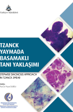
Granülpmatöz Hastalıklarda Tanısal Yaklaşım
Özet
Granülomatöz dermatit için karakteristik sitolojik bulgular, granülom yapıları ve üç farklı şekilde, yani
Langhans, yabancı cisim ve Touton tipinde görülebilen multinükleer dev histiyositlerdir. Granülom yapısı ve dev hücre pozitif ise granülomatöz dermatitin bakteriyel, fungal ve paraziter nedenleri araştırılmalıdır. Bu enfeksiyöz ajanlar yoksa nonenfeksiyöz granülomatöz hastalıklar açısından yabancı cisim, müsin yapısı, nekrobiyotik materyal ve köpüksü histiyositler incelenmelidir.
Anahtar Kelimeler: Granülom, kutanöz leishmaniasis, yabancı cisim granülomu, sarkoidoz
×
Kaynak Göster
Referanslar
- Durdu M, Baba M, Seçkin D. More experiences with the Tzanck smear test: cytologic findings in cutaneous granulomatous disorders. J Am Acad Dermatol 2009; 61: 441-50. [Crossref]
- Rahman S, Bari A. Laboratory profile in patients of cutaneous leishmaniasis from various regions of Pakistan. J Coll Physicians Surg Pak 2003; 13: 313-6.
- Dar NR, Khurshid T. Comparison ofskin smears and biopsy specimensfor demonstration of Leishmania tropica bodiesin cutaneousleishmaniasis. J Coll Physicians Surg Pak 2005; 15: 765-7.
- Haque A. Special stainsise in fungal infections. [Link]
- Arenas R, Moreno-Coutiño G, Welsh O. Classification of subcutaneous and systemic mycoses. Clin Dermatol 2012; 30: 369-71. [Crossref]
- Ilkit M, Durdu M, Karakaş M. Majocchi's granuloma: a symptom complex caused by fungal pathogens. Med Mycol 2012l; 50: 449-57. [Crossref]
- Hussein MR. Mucocutaneous Splendore-Hoeppli phenomenon. J Cutan Pathol 2008; 35: 979-88. [Crossref]
- Kojima M, Kashiwabara K, Itoh H, Masawa N, Miyawaki S. Imprint cytology of hepatosplenic suppurative candidal granuloma complicating acute leukaemia: three case reports. Diagn Cytopathol 2009; 37: 705-6. [Crossref]
- van Burik JA, Colven R, Spach DH. Cutaneous aspergillosis. J Clin Microbiol 1998; 36: 3115-21. [Crossref]
- Powers CN. Diagnosis of infectious diseases: a cytopathologist's perspective. Clin Microbiol Rev 1998; 11: 341-365. [Crossref]
- Durdu M, Güran M, Ilkit M. Epidemiological characteristics of Malassezia folliculitis and use of the May-Grünwald-Giemsa stain to diagnose the infection. Diagn Microbiol Infect Dis 2013; 76: 450-7. [Crossref]
- Woods GL, Walker DH. Detection of infection or infectious agents by use of cytologic and histologic stains. Clin Microbiol Rev 1996; 9: 382-404. [Crossref]
- DiCaudo DJ. Coccidioidomycosis: a review and update. J Am Acad Dermatol 2006; 55: 929-42. [Crossref]
- Civila ES, Bonasse J, Conti-Díaz IA, Vignale RA. Importance of the direct fresh examination in the diagnosis of cutaneous sporotrichosis. Int J Dermatol 2004; 43: 808-10. [Crossref]
- Vásquez-del-Mercado E, Arenas R, Padilla-Desgarenes C. Sporotrichosis. Clin Dermatol 2012; 30: 437-43. [Crossref]
- Talhari S, Talhari C. Lobomycosis. Clin Dermatol 2012; 30: 420-4. [Crossref]
- Isa-Isa R, García C, Isa M, Arenas R. Subcutaneous phaeohyphomycosis (mycotic cyst). Clin Dermatol 2012; 30: 425-31. [Crossref]
- Torres-Guerrero E, Isa-Isa R, Isa M, Arenas R. Chromoblastomycosis. Clin Dermatol. 2012 Jul-Aug;30(4):403-8. [Crossref]
- Chavan SS, Reddy P. Cytological diagnosis of chromoblastomycosis. J Cytol 2013; 30: 276-7. [Crossref]
- Mayorga J, Barba-Gómez JF, Verduzco-Martínez AP, Muñoz-Estrada VF, Welsh O. Protothecosis. Clin Dermatol 2012; 30: 432-6. [Crossref]
- Lass-Flörl C, Mayr A. Human protothecosis. Clin Microbiol Rev 2007; 20: 230-42. [Crossref]
- Chandler FW, Kaplan W, Callaway CS. Differentiation between Prototheca and morphologically similar green algae in tissue. Arch Pathol Lab Med 1978; 102: 353-6.
- Estrada R, Chávez-López G, Estrada-Chávez G, López-Martínez R, Welsh O. Eumycetoma. Clin Dermatol 2012; 30: 389-96. [Crossref]
- Hemalata M, Prasad S, Venkatesh K, Niveditha SR, Kumar SA. Cytological diagnosis of actinomycosis and eumycetoma: a report of two cases. Diagn Cytopathol 2010; 38: 918-20. [Crossref]
- Afroz N, Khan N, Siddiqui FA, Rizvi M. Eumycetoma versus actinomycetoma: Diagnosis on cytology. J Cytol 2010; 27: 133-5. [Crossref]
- Farnandes H, D'souza CR, Shekhar JC, Marla NJ, Swethadri GK, Naik R. Cytodiagnosis of actinomycetoma. Diagn Cytopathol 2009; 37: 506-8. [Crossref]
- EL Hag IA, Fahal AH, Gasim ET. Fine needle aspiration cytology of mycetoma. Acta Cytol 1996; 40: 461-4. [Crossref]
- Kramer SC, Ryan M, Bourbeau P, Tyler WB, Elston DM. Fontana-positive grainsin mycetoma caused by Microsporum canis. Pediatr Dermatol 2006; 23: 473- 5. [Crossref]
- Gabhane SK, Gangane N, Anshu. Cytodiagnosis of eumycotic mycetoma: a case report. Acta Cytol 2008; 52: 354-6. [Crossref]
- Padilla-Desgarennes C, Vázquez-González D, Bonifaz A. Botryomycosis. Clin Dermatol 2012; 30:397-402. [Crossref]
- Katkar V, Mohammad F, Raut S, Amir R. Red grain botryomycosis due to Staphylococcus aureus--a novel case report. Indian J Med Microbiol 2009; 27: 370- 2. [Crossref]
- Welsh O, Vera-Cabrera L, Welsh E, Salinas MC. Actinomycetoma and advances in its treatment. Clin Dermatol 2012; 30: 372-81. [Crossref]
- Hemalata M, Prasad S, Venkatesh K, Niveditha SR, Kumar SA. Cytological diagnosis of actinomycosis and eumycetoma: a report of two cases. Diagn Cytopathol 2010; 38: 918-20. [Crossref]
- Chatterjee D, Dey P. Tuberculosis Revisited: Cytological Perspective. Diagn. Cytopathol. 2014. [Crossref]
- Kathuria P, Agarwal K, Koranne RV. The role of fine-needle aspiration cytology and Ziehl Neelsen staining in the diagnosis of cutaneous tuberculosis. Diagn Cytopathol 2006;34: 826-9. [Crossref]
- Mahajan VK. Slit-skin smear in leprosy: lest we forget it! Indian J Lepr 2013; 85: 177-83.
- Fazu ŞA. Lepra'da epidemiyoloji ve bakteriyolojik teşhis. Türkiye Klinikleri 1987; 7: 133-136.
- Fassina A, Olivotto A, Cappellesso R, Vendraminelli R, Fassan M. Fine-needle cytology of cutaneous juvenile xanthogranuloma and langerhans cell histiocytosis. Cancer Cytopathol 2011; 119:134-40. [Crossref]
- Barroca H, Farinha NJ, Lobo A, Monteiro J, Lopes JM. Deep seated congenital juvenile xanthogranuloma. Acta Cytol 2007; 51:473-6. [Crossref]
- Chung YE, Kim EK, Kim MJ, Yun M, Hong SW. Suture granuloma mimicking recurrent thyroid carcinoma on ultrasonography. Yonsei Med J 2006; 47: 748-51. [Crossref]
- Kito Y, Hashizume H, Tokura Y. Rosacea-like demodicosis mimicking cutaneous lymphoma. Acta Derm Venereol 2012; 92: 169-70. [Crossref]
- Seifert HW. Demodex folliculorum causing solitary tuberculoid granuloma. Z Hautkr 1978;53: 540-2.
- Dong H, Duncan LD. Cytologic findings in Demodex folliculitis: a case report and review of the literature. Diagn Cytopathol 2006; 34: 232-4. [Crossref]
- Gaafar SM. Autofluorescence in demodex canis. Am J Vet Res 1964; 25: 233-5.
- Fernández-Díez J, Magaña M, Magaña ML. Cutaneous amebiasis: 50 years of experience. Cutis 2012; 90: 310-4.
- Murakawa GJ, McCalmont T, Altman J, Telang GH, Hoffman MD, Kantor GR, Berger TG. Disseminated acanthamebiasisin patients with AIDS. A report of five cases and a review of the literature. Arch Dermatol 1995; 131: 1291-6. [Crossref]
- Kenner BM, Rosen T. Cutaneous amebiasis in a child and review of the literature. Pediatr Dermatol 2006; 23: 231-4. [Crossref]
- Beer K, Beer MS, Appelbaum D. Granuloma annulare masquerading as erythema multiforme. J Drugs Dermatol 2013; 12: 694-7.
- Cano Martínez N, Fernández-Antón Martínez C, Barchino Ortiz L, Lecona Echevarría M, Campos Domínguez M. Dermatomyofibroma mimicking granuloma annulare. Dermatol Online J. 2011; 17: 3. [Crossref]
- Jouary T, Beylot-Barry M, Vergier B, Paroissien J, Doutre MS, Beylot C. Mycosis fungoides mimicking granuloma annulare. Br J Dermatol 2002; 146: 1102-4. [Crossref]
- Mehrota R, Dihingra V. Cytological diagnosis of sarcoidosis revisited: a state of the art review. Diagn Cytopathol 2011; 39: 541-8 [Crossref]

