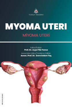
Uterus Anatomy
Selma Çalışkan
Ankara Yıldırım Beyazıt University, Faculty of Medicine, Department of Anatomy, Ankara, Türkiye
Çalışkan S. Uterus Anatomy. Yavuz AF, ed. Myoma Uteri. 1st ed. Ankara: Türkiye Klinikleri; 2025. p.1-6.
ABSTRACT
Uterus is a muscular organ containing an internal cavity. Upper segment of uterus comprises fundus and body while lower segment consists of isthmus and cervix. Uterus is an anteverted and anteflexed organ. It is intraperitoneal except its cervical part. Peritoneum reflects onto urinary bladder from uterus forming vesicouterine pouch and to rectum forming rectouterine pouch. From outer to inner surface, the uterus is composed of three layers: the perimetrium, myometrium, and endometrium. Müllerian ducts give rise to female reproductive system in fetal life. Improper fusion or resorption of Müllerian ducts may lead a series uterine anomalies. Uterus is held in normal position by true ligaments (fibrous tissue) and false ligaments (peritoneal folds). The endopelvic fascia covering the muscular floor of the pelvis (levator ani, obturator internus, piriformis, and coccygeus muscles) condenses to form pel- vic ligaments, which suspend the pelvic organs from the pelvic wall: cardinal ligaments (also known as transverse cervical or Mackenrodt?s ligaments), uterosacral ligaments, and pubocervical ligament. Uterine arteries and ovarian arteries supply uterus. Anterior trunk of internal iliac artery gives rise to uterine artery below the origin of obturator artery. Uterine artery gives an ascending tortuous branch which soon anastomosis with ovarian artery in the region of ovarian hilum. Descending branch of uter- ine artery supplies cervix and anastomoses with vaginal artery Uterus is predominantly innervated by sympathetic system. Sympathetic activity provides uterine contraction and vasocontruction. Efferent presynaptic sympathetic axons innervating uterus are derived from neurons in lateral horn of T12, L1 and L2 segments of spinal cord. Parasympathetic fibers in pelvic plexus responsible of uterine relax- ation and vasodilatation are derived from S2-4 segments of spinal cord.
Keywords: Uterus; Anatomy; Leiomyoma
Kaynak Göster
Referanslar
- Standring S. Gray's Anatomy. The Anatomical Basis of Clinical Practice. 41st ed. London; UK: Elsevier; 2016. p.1288-1300.
- Gilroy AM. Anatomi Temel Ders Kitabı.1.Baskıdan çeviri. Ankara:Palme; 2015. p.200-204.
- Singh I. Textbook of Anatomy With Colour Atlas. 3rd ed. New Delhi:Jaypee; 2005. p.708-711.
- Moore KL. Clinically Oriented Anatomy. 7th ed. China: Lippincott Williams and Wilkins; 2013. p.361, 385-387.
- Sancak B. Fonksiyonel Anatomi. 13. Baskı. Ankara: ODTÜ; 2017. p. 302-306
- Tortora GJ. Principles of Human Anatomy. 14th ed.USA: Wiley; 2016. p.859-861.
- Liatsikos SA. Pathogenesis and Aetiology of Female Genital Malformations in Grimbizis GF. Female Genital Tract Congenital Malformations Longon:Springer; 2015. P.15-22, 248. [Crossref]
- Sadler TW. Langman's Medical Embriology. 11th ed. China:Lippincott Williams and Wilkins; 2010. P252-254
- Snell RS. Clinical Anatomy. 4th ed. USA: Lippincott Williams and Wilkins;2000. P.96-100
- Kahle. Color Atlas of Human Anatomy, Vol 3. Revised 5th ed. Germany: Thieme P.292-303
- Wancura-Kampik, Ingrid. Segmental Anatomy: The Key to Mastering Acupuncture, Neural Therapy, and Manual Therapy.1st ed. Elsevier, 2008. P.332-336

